Describe the Anatomy of the Breast
Anatomy Physiology of the Breast. In humans breast tissue begins to enlarge at puberty.
Anatomy Of The Breast Memorial Sloan Kettering Cancer Center
1 New York NY USA 21 Anatomy This overview is not meant to be comprehensive.

. The Anatomy and Physiology of the Breast. The mammary glands are made up of lobules milk-producing glandular structures and a system of ducts that transport milk to the nipple. Breast tissue can develop anywhere along the milk line.
Within these tissues are milk-producing cells. The amount of fat. He described ligamenta suspensoria which connected to the skin and muscle.
The upper outer quadrant is the site of most breast tumors. Breast tissue begins to originate by the fourth week of fetal life. The nipple anatomy is adjusted to support the function of the breastThey are surrounded by a pigmented circular region of skin called the areola which becomes even more pigmented and prominent during pubertyThe areola shows small punctual elevations on its surface which are produced by the many areolar glands.
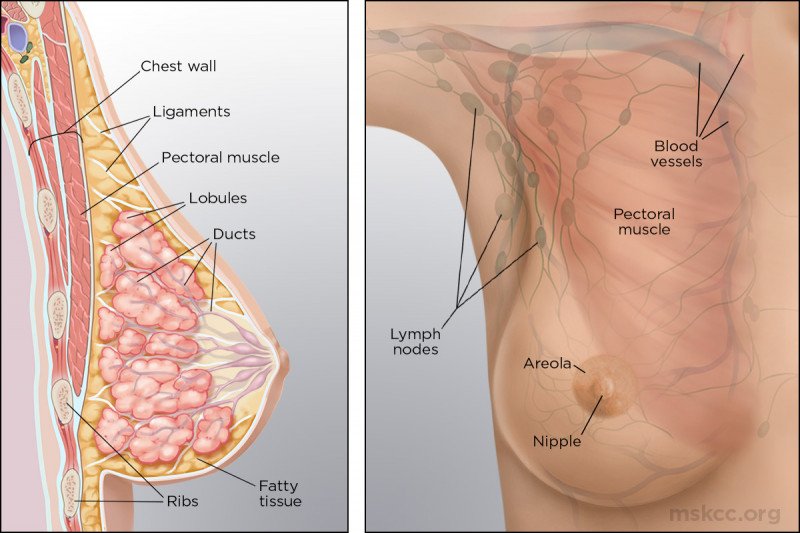
Rather it contains key factors that help provide a context in which to discuss breastfeeding. It grows along two ridges one on either side running from the armpit axilla to the groin. Figure 11 shows the different parts of the breast.
These are the so-called milk ridges or milk lines. Understanding the different parts and functions will help you better grasp the details of breast cancer. 1 Cooper wrote concerning the fascia mammae that it contained two layers one anterior to the breast gland and one posterior to it.
Areolas surround nipples with sebaceous glands Glandular Tissue Fibrous Tissue suspensory ligaments Adipose tissue Broken up into four quadrants. The authors set out to discover and describe a theory of superficial fascia structures responsible for breast shape. Knowing your body helps you to.
It extends horizontally from the lateral border of the sternum to the mid-axillary lineVertically it spans between the 2nd and 6th costal cartilagesIt lies superficially to the pectoralis major and serratus anterior muscles. Fibrous or supportive or connective tissue is the same tissue that ligaments and scar tissue are made of. Be aware of anything unusual.
New imaging techniques show. The breast anatomy of males and females is slightly different. All genders can get breast cancer.
Doctors refer to all non-fatty tissue as fibroglandular tissue. The breasts comprise glandular connective and fatty tissue. The structure of the breast.
Surrounding the nipple is the areola an area of darker skin that becomes both larger and darker during pregnancy. Located between the second and 6th rib. As you learn about breast cancer we will repeatedly reference the anatomy of the breast.
Areola surrounds nipple with sebaceous glands montgomery glands- secretes protective lipid material Glandular Tissue Fibrous Tissue suspensory ligaments Adipose tissue. Breasts are made up of fat and breast tissue along with nerves veins arteries and connective tissue that helps hold everything in place. Have a better dialogue with your doctor.
In women the breasts are composed principally of specialised tissue glandular tissue that produces milk. The main chest muscle the pectoralis muscle is found between the breast and the ribs in the chest wall. Tiny openings in the nipple allow milk to flow.
The female breasts or mammary glands are the modified appendages lying on the anterior chest wall. It is present in males as well but in most men it is not as prominent as it is in females. Tail of Spence in the axillary.
The female breast has a role in both nourishing the offspring as well as offering immunological. Women are more prone to benign noncancerous breast disease. The production of milk for lactation breast feeding.
The epithelial component of the tissue consists of lobules where milk is made which connect to ducts that lead out to the nipple. There are many books that describe breast anatomy and changes during pregnancy and lactation for those who would like to delve more fully into. The remaining part is made up of fatty tissue.
Fewer milk ducts average 9. Picture of the anatomy of the breast. Breast milk also contains bioactive factors that augment the infants immature immune system providing protection against infection and other factors that help digestion and.
Correlate changes in the female breast with. The breast is located on the anterior thoracic wall. Glandular tissue includes the breast lobes and breast ducts.
The breast is the tissue overlying the chest pectoral muscles. The incomplete breast consists mostly of adipose tissue but also lactiferous un. Lymphatic vessels in the breast drain excess fluid.
Review breast changes that occur during pregnancy. Describe the anatomy of the breast. The breasts are medically known as the mammary glands.
Relate the anatomy of the breast lymphatic system Review breast development in. Female breasts have milk ducts and glandular tissue that aid breastfeeding. Womens breasts are made of specialized tissue that produces milk glandular tissue as well as fatty tissue.
Female Breast Anatomy Function Parts and Pictures. Lymphatic drainage of the breast Functional drainage of the breast. Male and female breast nipples have many nerves that enhance sexual arousal.
The mature breast is located within the anterior thoracic wall lying atop the pectoralis major muscle. Breast milk contains all the nutrients that an infant needs in the first 6 months of life including fat carbohydrates proteins vitamins minerals and water 1234. It is easily digested and efficiently used.
The medical name for breast is mammary gland. Most 75-90 of the lymphatic drainage of the breast is to the ipsilateral same side axillary nodesNearly all lymphatics of the breast drain along a subdermal plane into the axillae typically collecting in a single sentinel lymph node at the lateral border of the pectoralis major muscle. The nature of the superficial fascia system that surrounds the breast and its attachments to the chest were studied in 12 cadaver breast dissections and in clinical cases of both cosmetic and reconstructive breast.
Pubertal changes lead to incomplete development of the breast a process which is only completed during pregnancy. It is quite common to have breast tissue up toward and even in the armpit. Each breast consists of tissue overlying the chest wall muscles the pectoral muscles.
They are located between the second and 6th rib. The breast is an organ whose structure reflects its special function. Describe the anatomy of the breast.
In 1840 Sir Astley Cooper published his book On the Anatomy of the Breast. Fatty tissue fills in the spaces between glandular and fibrous tissue and largely determines your breast size. Breast growth begins at puberty in humans in contrast to other.
Range 4-18 Milk ducts branch closer to nipple 65 of glandular tissue within 30mm radius from base Milk ducts are small and do not display lactiferous sinuses dilated portion beneath nipple-only dilate with milk ejection reflex. Relate the anatomy of the breast lymphatic system. Describe the procedure for teaching breast self-examination and incorporating health promotion concepts when performing an assessment of the breasts.

Breast Anatomy Breast Cancer Breastfeeding Conditions
Breast Anatomy Breast360 Org The American Society Of Breast Surgeons Foundation
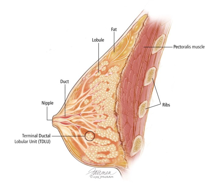
Overview Of The Breast Breast Pathology Johns Hopkins Pathology


Comments
Post a Comment